how do they x ray babies hips
Lying on his back one leg in the knee bends at an angle. You will lay face up on an X-ray table.

Infant Diagnosis International Hip Dysplasia Institute
Around 6 months of age enough bone is present in an infant hip to make an X-ray more accurate than.

. An X-ray of the pelvis focuses specifically on the area between your hips that holds many of your reproductive and digestive organs. A hip X-ray is a safe and painless test that uses a small amount of radiation to make images of the hip joints where the legs attach to the pelvis. This image shows the soft tissues and the bones of the pelvis and hip joints.
It is put on by an orthopedic surgeon while using x-ray to make sure the hip is aligned correctly. The X-ray image is black and white. Its a cast that goes around both hips and down the leg to keep the hips aligned.
The maximum visual information is given by the x-ray of the hip joint in two projections. How do they x ray babies uk are a topic that is being searched for and liked by netizens today. Often a special baby xray tube is used to hold the child still and capture sharper.
You will go in the room with him he will need to. X-rays of the hip can reveal bone tumors and diagnose bone cancer. Your healthcare provider may use hip X-rays to diagnose and treat health conditions involving your hips.
The picture shows the inner. An X-ray technician will take pictures of the hip. It might help to feed your baby just before the ultrasound to make your little one more relaxed.
The exact procedure will depend on the type of scan needed. You will go in the room with him he will need to be stripped from the waist down they will take x-rays of him flat on his back legs dead straight and together you wil be able to. Its a very effective way of looking at the bones and can be used to help detect a range of conditions.
For a simple baby hand xray the child can usually be scanned using a standard device using just a light restraint. Hip X-rays are quick easy and painless procedures. From the front anteroposterior view or AP from the side lateral.
You will go in the room with him he. Two tests are performed called the Barlow. If an X-ray of the hip joints is performed according to Launstein Lauenstein then the patients position looks like this.
A hip X-ray is a safe and painless test that uses a small amount of radiation to make images of the hip joints where the legs attach to the pelvis. This is also why most doctors want. An X-ray technician will take pictures of the hip.
If it persists they may put on a spica cast. An X-ray of the hip joints in children is carried out according to strict indications - only after the child reaches nine months. A hip X-ray is a test that produces an image of the anatomy of your hip.
The reasons for this are unknown but this is a reason why some doctors insist on prolonged bracing even when the x-ray or ultrasound seems normal.

Pediatric Hip Disorders Radsource
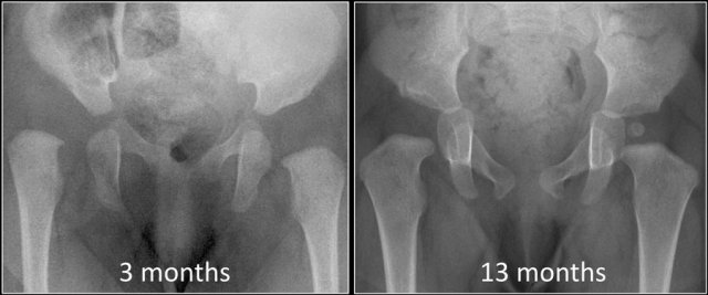
The Radiology Assistant Developmental Dysplasia Of The Hip
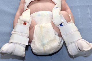
Developmental Dysplasia Of The Hip Nhs
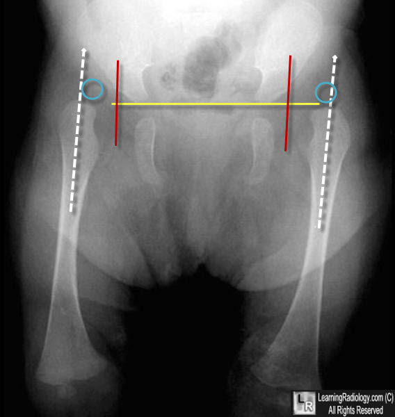
Learningradiology Developmental Dislocation Dysplasia Of The Hip
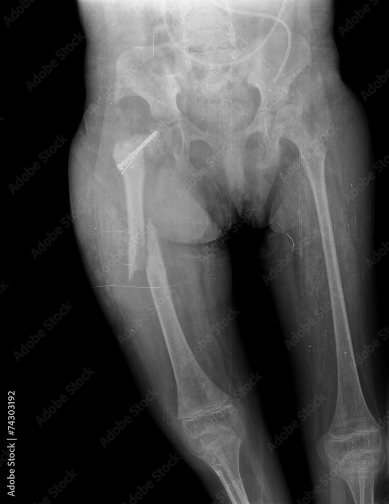
X Ray Of Child Hip And Broken Leg Stock Photo Adobe Stock

X Ray Exam Pelvis Connecticut Children S
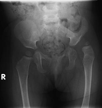
Hip Dysplasia In The Child And Adolescent Musculoskeletal Key
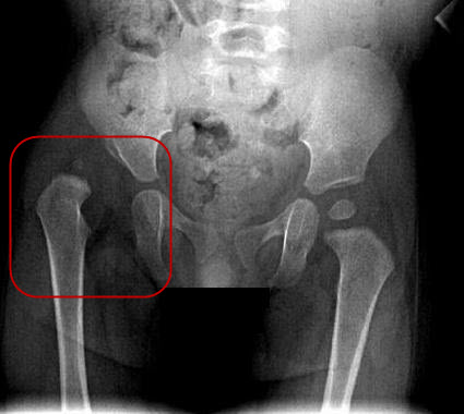
X Ray Screening International Hip Dysplasia Institute

Radiology In Ped Emerg Med Vol 5 Case 12

Pediatric Pelvis Ap View Radiology Reference Article Radiopaedia Org

Other Resources For Parents International Hip Dysplasia Institute
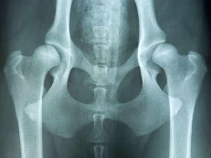
Congenital Hip Dislocation Causes Symptoms And Diagnosis

Pediatric Hip Disorders Radsource
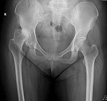
X Ray Of Hip Dysplasia Wikipedia
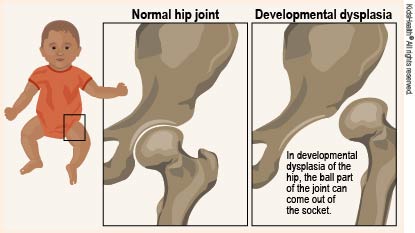
Developmental Dysplasia Of The Hip For Parents Nemours Kidshealth
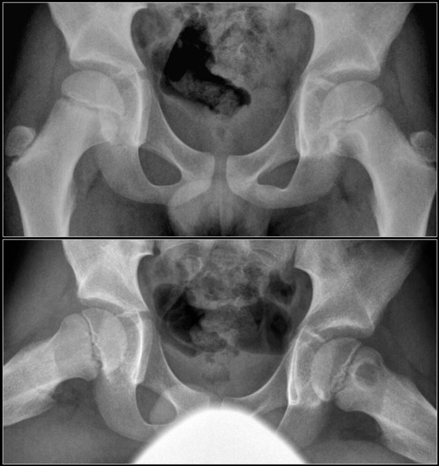
The Radiology Assistant Hip Pathology In Children

Infant Diagnosis International Hip Dysplasia Institute

Radiograph Of The Pelvis Of A Five Month Old Infant Showing The Download Scientific Diagram
